Custom/ Other (IHC, ISH, etc.)
- ELISA/ EIA (Enzyme Linked Immuno-absorbent Assay)
Please call for more information about PDL offered ELISA/ EIA's.
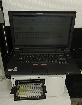
The presence or absence of antibodies in serum or plasma can be determined qualitatively by Enzyme Immuno Assay. In these assays, microtiter plate wells are coated with antigen (typically purified viral lysate). Diluted serum is incubated in the wells. If specific antibody is present it will bind to the antigen. The enzyme conjugated detector antibody is added next. Finally a chromogen reagent is added. The development of color in the wells signifies the presence of specific antibody.
The minimum sample requirement is 1 ml of serum or plasma sent frozen.
- Immunohistochemistry/in situ hybridization (IHC/ISH)
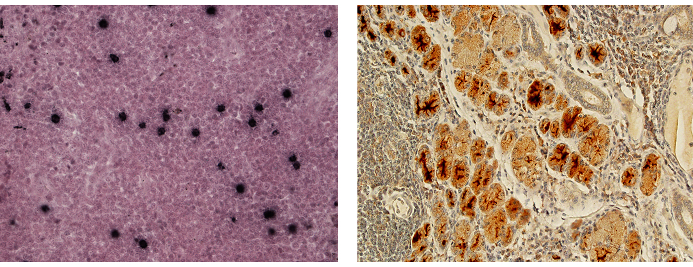
Immunohistochemistry, or IHC, refers to the process of visualizing antigens (e.g. proteins) in cells of a tissue section. Specific antibodies are used to bind specifically to antigens in biological tissues. Immunohistochemical staining is widely used in the diagnosis of abnormal cells such as those found in cancerous tumors. Specific molecular markers are characteristic of particular cellular events such as proliferation or cell death (apoptosis). Virus proteins can be visualized in specific tissues and specific cell compartments using this technique. This is particularly useful in cases where suspicion of the infection doesn't occur until after euthanasia and typical blood and plasma samples are not available.
Immunohistochemistry stains thin slices of formaldehyde-fixed paraffin-embedded (ffpe) tissues on glass slides. Specific antibody (poly- or monoclonal) targeted to the antigen is incubated with the tissue. A secondary antibody, conjugated to an enzyme (e.g. peroxidase) or fluorescent tag is then used to visualize the binding. The enzyme/conjugate catalyzes a reaction that results in colored deposition in the tissue. PDL offers SRV IHC using a monoclonal gp20 antibody that cross-reacts with serotypes 1-5. Please inquire about custom IHC for other pathogens.
In situ Hybridization, or ISH, refers to the process of visualizing specific nucleotide sequences in tissue sections. The tissue is treated to encourage hybridization. A conjugated probe is used to specifically hybridize to the sequence of choice and then visualized. We are currently validating ISH for SRV and other pathogens. This assay is offered on a custom basis.
- CUSTOM / OTHER TESTS
Other assays are available or can be developed to answer specific research or diagnostic questions. The charge is actual labor plus cost of reagents. Please contact us to discuss.
Some examples include:
- Virus isolation for SIV, SRV, SFV, RhCMV and RRV
- SRV Immunohistochemistry (IHC)
- SRV In-situ Hybridization (ISH)
- PCR typing for SRV 1-5
- Quantitative PCR
- RNA PCR
- Quantitative antibody titers
- Cytokine /Chemokine Detection and Quantitation Assays (EIA and MIA) including IL1b, IL1Ra, IL2, IL4, IL5, IL6, IL8, IL10, IL12 (p70), IL13, IL15, IL17, IL18, sCD40L, G-CSF, GM-CSF, IFNg, MCP-1, MIP-1a, MIP-1b, TGFa, TNFa, VEGF, Rantes, TGF-1
- Custom culturing and processing.